A camera placed below the gumline can find calculus that instruments leave behind. The finding changed how one periodontal practice trains and uses its hygienists.
What feels like a smooth root may still carry deposits. Once subgingival calculus breaks apart, fractured pieces can stay attached to the root. This happens more often in pockets deeper than about 4 mm, where tactile instrumentation becomes less effective.
Stambaugh and colleagues found that complete removal of plaque, calculus and altered cementum occurred at an average depth of only 3.73 mm.²
A 2026 review reported that guideline-based treatment for stage III-IV periodontitis reached its clinical targets in 93.3% of treated sites and in about half of patients. Outcomes were less favorable around posterior teeth, in deeper pockets and where furcation involvement was present.
Root cleanliness is the treatment goal. Current clinical guidance still places subgingival mechanical instrumentation at the center of care. Clinicians may call it deep cleaning, root-surface debridement or scaling and root planing. The work removes calculus and biofilm, reduces inflammation and creates conditions for tissue healing.
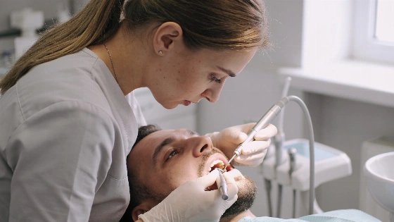
The practice starts with 24% EDTA gel on the root surface. A microbrush spreads the gel for one minute. An irrigating syringe then rinses it away. The periodontal endoscope comes next. It helps the clinician locate and remove calculus with ultrasonic and hand instruments.
After the visual check, the clinician burnishes the root with 24% EDTA for another minute. The surface is then rinsed thoroughly.
The sequence targets deposits that conventional scaling and root planing can miss. In an extracted-tooth study, conventional instrumentation used ultrasonic devices, new Gracey curettes and 3.5x loupes. Residual calculus remained across 45% to 53% of the test area, even after clinicians reached a clinical endpoint of no detectable calculus.
Periodontal research published in 2026 continues to examine clinical outcomes after subgingival instrumentation, including prospective observations of inflammatory markers after treatment. The continuing research activity shows that root-surface debridement remains an active clinical question rather than a settled technical detail.
The endoscope added more than magnification. It challenged a long-held clinical assumption. Current 2026 clinical materials describe the same periodontal aim: a clean root surface lets gingival tissue reattach closer to the tooth and can help reduce pocket depth.
The practice first moved toward hygienist-led periodontal care after a period of study with Dr. Perry Ratcliff in Scottsdale, Arizona. Ratcliff used retraction cord, citric acid and tetracycline to improve access and visibility.
When the approach reached a surgical periodontal practice, the clinician stopped routine periodontal surgery for eight months. Only four isolated periodontal surgical procedures took place during that period. Patients who needed surgery received it at no charge.
Thirteen years later, periodontal endoscopy supplied the missing visual check. Hygienists who saw calculus beneath apparently clean surfaces were often embarrassed by what they had missed. The more useful lesson was clear: a smooth surface detected with a no. 17 explorer does not prove root cleanliness.
Three hygienists at the practice now use the endoscope to visualize clean surfaces and reassess the root.
Cleaning alone does not complete the periodontal diagnosis. The practice identifies three healing needs: root cleanliness, control of aggressive microorganisms when they contribute to disease, and stable occlusion. Routine mouth rinses cannot replace professional subgingival instrumentation. They do not remove calculus or mature biofilm attached below the gumline.
For suspected rapid destruction, the clinician looks for a periodontal probe that drops into the pocket without resistance. The root must also show no detectable calculus. These findings appear in fewer than 10% of cases in the reported practice.
When they do appear, the protocol calls for salivary testing. The patient then receives an antibiotic regimen, usually amoxicillin and metronidazole. Reassessment takes place one week after the antibiotics are completed. Clinical improvement supports the next step, periodontal endoscopy.
Occlusion is the third checkpoint.
Fremitus means that a tooth moves when the patient is in occlusion. In a periodontally compromised tooth, that movement can limit healing. The reported response is bite adjustment. The clinician may remove some tooth structure from the compromised tooth or add composite to adjacent teeth so the load is shared.
This approach fits the wider clinical lesson in an earlier case analysis of tooth movement linked to periodontal bone loss. Disease control must come before clinicians declare a tooth beyond saving.
Current treatment benchmarks set clear limits. Pockets deeper than 4 mm that continue to bleed have not reached the desired clinical endpoints. The same applies to pockets measuring 6 mm or more.
Too many teeth are labeled periodontally hopeless before clinicians address the root surface, microorganisms and occlusal load together. The reported evidence supports a stricter standard. Visualize what can be seen. Clean what remains. Address the biological and mechanical causes before choosing extraction or surgery.
That puts the dental hygienist at the center of periodontal care, not at its edge.
According to the 2026 clinical guidance summarized by BestDentalTourism, root-surface debridement is not simply about making the root feel smooth. Its purpose is to reduce inflammation and create a biologically favorable surface for healing. The distinction keeps visual assessment, reassessment and maintenance tied to the patient's long-term periodontal stability.