
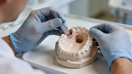
After a tooth is removed, the socket's remaining bone walls shape how much ridge resorption follows.
A socket graft may preserve some ridge width and height, but current evidence does not show that routine grafting improves implant survival. The clinical question is which sites need that dimensional change to support the planned treatment.
That distinction challenges calls to graft every extraction site except third molars. A four-wall socket with adequately thick bone may heal without a graft. A thin or damaged buccal plate can put later implant treatment at greater risk. The aim is to match socket preservation to the anatomy and clinical need.
What the evidence establishes
The 2021 Cochrane review included 16 randomised controlled trials involving 426 adults and 524 extraction sites. Compared with extraction alone, xenograft procedures were associated with 1.18 mm less ridge-width loss and 1.35 mm less height loss on average. Cochrane rated the evidence for these dimensional benefits very uncertain.
The review found no demonstrated difference in the need for additional augmentation or in implant failure. Its confidence intervals were wide, so the findings do not establish that outcomes are equivalent. None of the included trials reported aesthetic or prosthodontic outcomes.
A smaller measurement change may not translate into a meaningful clinical benefit.
Studies commonly measure ridge width or height, rather than whether the remaining bone allows an implant to be placed in the required position. A systematic review by Avila-Ortiz and colleagues reported absolute differences of 1.89 mm in buccolingual width and 2.07 mm in mid-buccal height for non-molar teeth. Those averages do not show whether a particular site will need later augmentation.
A later randomised trial by the same group found that additional augmentation was considered necessary in 48.1% of extraction-only sites, compared with 11.5% of sites treated with alveolar ridge preservation. That gap contrasts with the less conclusive Cochrane result. In both trial groups, baseline buccal bone thickness strongly predicted resorption, putting anatomy at the centre of treatment selection.
The socket itself matters
Remaining bone walls influence socket healing. Araújo and colleagues reported less resorption in four-wall sockets with adequate bone than in compromised sockets. They identified the number and thickness of the remaining walls as primary determinants. The most pronounced remodelling occurs early. Experimental work by Araújo and Lindhe described marked dimensional changes during the first eight weeks after extraction. In anterior maxillary sites, Chappuis and colleagues found that sockets with a buccal plate under 1 mm lost a median of 7.5 mm of mid-buccal height at eight weeks. Thick-phenotype sites lost 1.1 mm. Those figures apply to the anterior maxilla and should not be carried over wholesale to posterior teeth.
Whatever the grafting decision, preserving socket walls during extraction matters. So does thoroughly removing granulomatous or infected tissue. A graft cannot make up for avoidable damage to the bone that remains.
Graft materials are generally osteoconductive rather than osteogenic: they provide a scaffold, while bone-forming cells come from surrounding bone. Autogenous bone is the exception described in the source literature. Residual particles may be associated with reduced new bone formation or delayed remodelling. Reported complications include inflammation and pain, as well as membrane exposure, particle loss, infection, gingival recession and dry socket. The Cochrane review reported no serious adverse events in its included trials. These concerns therefore relate to morbidity and predictability, not evidence of serious harm.
Planning for the individual site
Clinicians are more likely to consider grafting when a socket has lost one or more walls, the buccal plate is thin, aesthetic demands are high or a draining fistula suggests a defect. Where CBCT imaging is already indicated, measuring buccal bone thickness can help inform the decision. For immediate implant placement, the gap between the implant and buccal bone also matters. Research reviews report greater benefit from grafting when that gap exceeds 2 mm. The 4th EAO Consensus Conference reported similar feasibility of implant placement after ridge preservation and unassisted healing, with comparable implant-related outcomes across different ridge-preservation protocols.
If clinically important bone loss becomes apparent later, guided bone regeneration at implant placement remains an established option. A classification published by the first Giuseppe Cardaropoli Foundation Consensus Conference also includes spontaneous healing followed by early implant placement, with contour augmentation if needed, among preferred or alternative options for several socket classes.
Patients should be told before extraction that later grafting may be needed. Clinicians who do not place the implant themselves can check healing with the implant provider and use the result to calibrate future decisions. Perth proclaimed October 10, 2026, as National Dental Care Day, with prevention and access in focus.